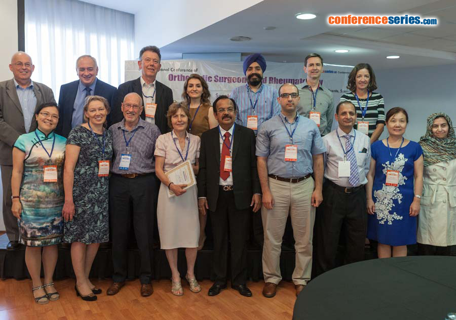
Manuel Villanueva
Avanfi Institute, Spain
Title: Ultrasound guided gastrocnemius recession: A new ultra-minimally invasive surgical technique
Biography
Biography: Manuel Villanueva
Abstract
Introduction: Gastrocnemius equinus is defi ned as ankle dorsifl exion<10º with the knee extended. Th e equinus deformity alters foot biomechanics, predisposing to conditions such as Achilles tendinosis, fl atfoot, diabetic foot ulcer, metatarsalgia, plantar fasciitis, midfoot arthritis and nerve entrapment. In children, the deformity has been associated with equinus foot, spasticity and cerebral palsy. Th erefore, gastrocnemius recession has many well documented indications. We present an ultrasound guided ultra-minimally invasive technique for gastrocnemius recession. Materials & Methods: In 22 cadavers we checked the technique was eff ective and safe. Th en we performed gastrocnemius recession in 23 patients (25 cases), 18 males and 5 females on an outpatient regimen. Mean age was 42 years (13-61). In 11 cases the indication for the procedure was non-insertional Achilles tendinopathy. In 5 patients, the indication was gastrocnemius retraction in the presence of plantar fasciitis. US guided Achilles tenotomy, release of the paratenon or selective plantar fasciotomy were combined with gastrocnemius recession. Th e age range of patients with Achilles tendinopathy or plantar fasciitis was 37-51 years. In 3 patients (4 cases) the indication was equinus foot. Th e ages were 13, 14 and 15 years. Ultrasound guided plantar fasciotomy was performed at the same time. In 5 patients (50-61 years) the indication was metatarsalgia and forefoot overloading with no hammertoe or any other forefoot condition. All patients had at least 6 months of failed conservative management prior to surgery. Surgical Technique: Th e instrument set included long needles (a 16 gauge, 1.7 mm diameter Abbocath), a V-shaped straight curette, a blunt dissector, a hook knife (Aesculap 2, 3 mm) and an ultrasound device (Alpinion ECube15) with a 10-17 MHz linear transducer and the Needle Vision Plus™ soft ware package. Th e patient is placed prone, under local anesthesia plus sedation without lower limb ischemia. Recession is performed via one or two incisions (1-2–mm each) positioning the instruments beneath the sural nerve. No stitches are required, just adhesive strips and elastic bandage. Active dorsifl exion and plantar fl exion of the ankle are encouraged immediately aft er surgery. Partial weight bearing is allowed the day of surgery, aided with crutches. Results: In the clinical series, pain, function and ankle dorsifl exion increased signifi cantly for every patient in the study (mean, 14º; STD 3º). VAS score improved from 7 (6-9) to 0 (0-1) and AOFAS score improved from a mean of 30 (20-40) to 93 (85-100), at 6 months. All athletes returned to their previous sports aft er 6 months. Superfi cial hematomas were common in the series and some patients developed internal hematomas (observed by ultrasound) at the areas of the tendon and muscle surrounding the recession until the third month. Th ere were no instances of over lengthening or Achilles tendon rupture, infections, wound or nerve complications. Discussion: Open or endoscopic gastrocnemius lengthening require epidural anesthesia, lower limb ischemia and stitches. Ultrasound guided ultra-minimally invasive gastrocnemius recession allows continuous visualization of nerves and vessels without ischemia. It can be combined with other US guided ultra-minimally invasive techniques (plantar fasciotomy, Achilles tenotomies) to ensure minimal pain with excellent outcomes and no signifi cant morbidity.


